Troubling Weight Trend

The latest obesity statistics demonstrate that too many Americans continue to slide down a slippery slope toward a poorer quality of life and many otherwise preventable conditions like heart disease, stroke and diabetes. Thirty-nine states now have 25% or more of their population classified as obese.

As recently as twenty years ago, not one state had over 15% obesity, and now the lowest rate is 19.8% in Colorado, the only state currently under twenty percent. Mississippi continues to hold the top position, with 34.4% of their citizens considered obese. These number don’t even count those who would be thought of as “overweight,” but only those who meet the clinical definition of obesity, having a body mass index of 30 or more. (To find your body mass index, or BMI, divide your weight in kilograms by your height in meters squared — it roughly translates to about thirty pounds or more overweight, or someone five-foot-nine weighing 203 pounds or more.)

It is time to change this scary trend! For more information on how our natural strategies can help call 565-7474 or visit www.nevadachiro.com.


Enhanced by Zemanta

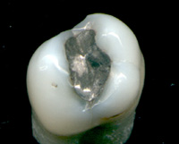
Silver Fillings Finally Being Regulated

An amalgam used as a restorative material in a...
Image via Wikipedia

The U.S. Environmental Protection Agency (EPA) has announced it will regulate the mercury used in dental amalgam fillings.

Responding to concerns about mercury amalgam pollution in the environment, the pro-mercury American Dental Association (ADA) had lobbied for “voluntary” self-regulation for its members. However, since the ADA’s position (and that of nearly all its more than 155,000 dentist members) is that mercury amalgams pose no environmental threat, self-regulation would have done little or nothing to stop dentists from continuing to dump tons of mercury into our environment.

Mercury released by amalgam-using dentists pollutes our water, our air, and our land, resulting in devastating environmental health effects. According to the EPA, 50 percent of all mercury entering local waste treatment plants comes directly from dental offices, where millions of bits and pieces of amalgam fillings are sent down the drain every year.

But the EPA explained in its announcement about regulating mercury amalgam that once dental mercury enters the environment, “certain microorganisms can change elemental mercury [which comprises about 50% of dental mercury amalgam) into methylmercury, a highly toxic form that builds up in fish, shellfish and animals that eat fish. Fish and shellfish are the main sources of methylmercury exposure to humans. Methylmercury can damage children’s developing brains and nervous systems even before they are born.”

Another problem, say consumer groups against the use of mercury amalgams, is the indiscriminate polluting of the air via crematoria. Tens of thousands of people with mercury fillings are cremated every year, and significant amounts of toxic mercury are released into the atmosphere along with the smoke.

For decades, the EPA maintained what critics called a “midnight deal” with the ADA: a “memorandum of understanding” by which the ADA was actually in charge of environmental safety in dental offices through, for example, the use of devices for trapping dental mercury before it goes down the drain. Under this arrangement, these devices were either woefully inadequate or woefully underused. As a spokesperson for the Consumers for Dental Choice said, it was like putting Colonel Sanders in charge of the chicken coop.

Teamed with other environmental groups to protest the status quo and demand new regulations, the Consumers for Dental Choice, established in 1996 by consumer advocates, mercury poisoning victims, scientists and mercury-free dentists, helped organize a congressional hearing to address the failure of ADA’s voluntary approach and the ever-increasing problem of dental mercury pollution.

As a result of all the efforts taken by numerous groups over the years, the EPA says it finally will propose a rule to regulate dental mercury in 2011. The public can participate by submitting public comments before the rule is finalized in 2012.

Want information about how to combat the effects of mercury? Check out www.nevadachiro.com or call (702) 565-7474.

SOURCES: Environmental Protection Agency, September 2010, http://yosemite.epa.gov/opa/admpress.nsf/d0cf6618525a9efb85257359003fb69d/a640db2ebad201cd852577ab00634848

Enhanced by Zemanta

20 PERCENT OF CALIFORNIA HOSPITALS FAIL TO REPORT MEDICAL ERRORS

Nearly 90 California hospitals – more than 20 percent of 418 hospitals in the state required by law to report medical errors – have gone more than three years without reporting any errors at all, says the California Department of Public Health.

Considering the fact that a recent report in the Journal of the American Medical Association found no change in patient harm error rates over the past decade, it is pretty hard to believe these hospitals are error-free.

Health officials in the state apparently agree, and have instituted investigations into the 87 hospitals apparently claiming no significant mistakes in patient care. They are calling for immediate clarifications – either confirmation that the hospital is indeed error free, or full disclosures and reports of errors.

According to a report in the Los Angeles Times, patient advocates are saying the lack of reports is an indication that hospitals are unwilling to police themselves.

A spokesperson for the Santa Monica-based advocacy group Consumer Watchdog told the newspaper it is “almost inconceivable” that so many hospitals have been error-free for three years.

State Sen. Elaine Alquist (D-Santa Clara), who wrote the medical error law, told the LA Times she was concerned that errors are going unreported. “What are the chances that nearly a quarter of California’s hospitals didn’t have a single medication, surgical or safety error since the reporting requirement became law?” she asked.

California’s law governing error reporting lists 28 medical errors that hospitals must report. These are the types of errors that typically place patients at risk of death or serious injury. After investigating each error, the Health Department can levy fines of $50,000 for the first incident, $75,000 for the second and $100,000 for the third or subsequent error.

But even more money could potentially change hands. Hospitals have five days to file an error report, after which the state can levy up to $100 a day for each day the hospital fails to report. We are looking at three years of failing to report, which could add up to even more money the hospitals could have to find. That’s not an easy thing to do in the health care field, even in the best of times, and these are far from the best times.

Are you ready for holistic care? Check out www.nevadachiro.com or call (702) 565-7474.


SOURCE: Los Angeles Times, Nov. 27, 2010 http://articles.latimes.com/2010/nov/27/local/la-me-hospital-errors-20101128

Enhanced by Zemanta

Toxins from Cups

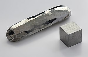
cadmium bar. Purity 99.999 %.
Image via Wikipedia

ARE YOUR KIDS DRINKING FROM TOXIC CUPS?

In June of 2010, the U.S. government recalled 12 million promotional drinking glasses at a fast food restaurant. The glasses contained cadmium — a toxic chemical that can cause a list of very nasty and dangerous conditions including:

* Emphysema

* Fatigue

* Headache

* Vomiting

* Anemia

* Lack of sense of smell

* Kidney dysfunction

* Reduced bone mineral density

* Neuropsychological impairment

* Increased risk of prostatic cancer

Cadmium serves no purpose in the human body, is toxic at very low levels, and once it gets in there, it’s hard to get rid. But it’s great for making those vivid images on the cups kids clamor for, even collect, in some fast food restaurants.

Cadmium is just one of the many toxic chemicals our kids are exposed to every day. Lead is another one. You would think we would have gotten rid of lead in kids’ products long ago, but the U.S. government has recalled more than 17 millions toys containing high levels of lead in just the last three years.

The Consumer Product Safety Commission also banned the use of phthalates — the chemicals used to make hard plastic pliable — in kids’ products in 2008. Some phthalates are banned, others are still under investigation and still being used. They, too, may be banned.

However, the wheels of government regulation turn slowly. And if 12 million cups and 17 millions toys can get out there, we each need to take action to prevent exposure.

To be safe, avail yourself of the many manufacturers that guarantee their products do not contain toxic chemicals. Give your kids a healthy start in life.

SOURCE: Science Daily: http://www.sciencedaily.com/releases/2010/11/101110123941.htm

Have questions concerning this information? Check out www.nevadachiro.com or call 565-7474.

Enhanced by Zemanta

How Posture Improves Athletic Performance

At sea aboard USS Bataan (LHD 5) Sept. 24, 200...
Image via Wikipedia

As an athlete, your regimen probably consists of the usual exercises: cardiovascular endurance, strength training, aerobic conditioning, etc.  But while these workouts may aid in improving your overall performance, a new study has shown that athletes are overlooking a vital component of a successful training regimen: maintaining excellent posture.

A 2004 study conducted by the American Medical Athletic Association (AMAA; www.amaasportsmed.org) concluded that athletes who want to optimize their performance should focus on their posture.  The study reported that “postural repositioning exercises, breathing exercises, gluteal, hamstring and abdominal/oblique exercises alone can provide for improved…performance as a result of improved posture.”  The study also concluded that improved posture can alleviate aches and pains, as well as prevent common sports injuries.

So why does good posture play such an important role in how well you’ll run during the big marathon, football game or at your next tennis match?  Simple: it’s because improper application of pressure on your spine, joints, muscles and nerves can all contribute to a less-than-optimal athletic performance.

Think about it in terms of a car.  No matter how much top-notch oil you pour into it, or how powerful the engine is, if the fundamental mechanics of the car aren’t properly aligned or working, you won’t get the maximum performance out of the car.  The human body functions in the same way: even if your diet and exercise routines are highly optimized for an Olympic outcome, you won’t get the expected results if your body has been strained by poor posture.

So what exercises can you do to improve your posture?

Experts recommend that all athletes include yoga and meditative practices in their training regimen as a way of strengthening key muscles and joints.  While yoga teaches how to find and maintain excellent posture, meditative sessions can reinforce proper breathing techniques, which can also relieve strain on muscles and joints.  Try this simple exercise, known as “belly breathing”:  breathe in through the nose and into the diaphragm for a count of five, and then exhale by “sighing” for a count of seven.  By using this breathing technique, athletes will not only relieve stress to the ribs, shoulders and backs – this type of breathing also requires less energy to use.   And as any athlete knows, less energy expelled on breathing means better performances.

Many athletes may require more assistance with corrective posture to enhance their athletic performance, which is where our combination of physical rehabilitation and chiropractic services help.  At an initial consultation, the athlete’s overall balance, structure and tension will be assessed to determine the root cause of poor posture.  Once the mechanical or structural problem has been identified, a custom program will be developed to help the athlete correct the imbalance and regain full range of athletic ability and performance.

When considering treatment for correcting posture related problems and enhancing athletic performance, our clinics in Henderson NV comes highly recommended by physicians in Clark County.  Feel free to contact us for a Sports Performance Evaluation, by calling us at (702) 565-7474 or at www.nevadachiro.com.

Enhanced by Zemanta

The Facts about Gastroesophageal Reflux Disorder

I wanted to share this article by one of my mentors, Dr. Howard Loomis. He is the godfather of enzyme replacement therapy.

by Howard Loomis, D.C.

Before beginning this discussion, I wish to point out that current diagnosis of this seemingly pandemic condition is done without physical examination or objective testing. Recommendation of antacids, proton-pump inhibitors, and histamine antagonists, thus canceling normal digestion in the stomach, can only lead to chronic degenerative conditions.

GERD is a condition that is described as occurring when the acidified liquid contents of the stomach back up into the esophagus. This includes not only stomach acid, but pepsin as well and may even include bile that has backed-up into the stomach from the duodenum. The liquid can inflame and damage the lining of the esophagus, if it is not protected by the mucosal lining.  This is in and of itself not a disease.

The symptoms of uncomplicated GERD are heartburn, regurgitation, and nausea, but these symptoms only occur in a minority of patients. This statement requires some explanation.

Reflux of the stomach’s liquid contents into the esophagus occurs in most normal individuals. In fact, one study found that reflux occurs as frequently in normal individuals as in patients with a diagnosis of GERD. There are several mechanisms that prevent the symptoms from occurring:

  • Most reflux occurs during the day when we are upright and refluxed liquid is more likely to flow back down into the stomach due to the effect of gravity.
  • When awake we repeatedly swallow and each swallow carries any refluxed liquid back into the stomach. Also saliva contains bicarbonate and with each swallow it travels down the esophagus and neutralizes the small amount of acid that remains in the esophagus after gravity and swallowing have removed most of the liquid.
  • At night while sleeping, gravity is not in effect, swallowing stops, and the secretion of saliva is reduced. Therefore, reflux that occurs at night is more likely to result in acid remaining in the esophagus longer and can cause damage to the esophagus.

Symptoms of Uncomplicated GERD

Heartburn
When acid refluxes back into the esophagus in patients with GERD, nerve fibers in the esophagus are stimulated. This nerve stimulation results most commonly in heartburn, the pain that is characteristic of GERD. Heartburn usually is described as a burning pain in the middle of the chest. It may start high in the abdomen or may extend up into the neck. In some patients, however, the pain may be sharp or pressure-like, rather than burning. Such pain can mimic heart pain or angina. In other patients, the pain may extend to the back.

Episodes of heartburn tend to happen periodically. This means that the episodes are more frequent or severe for a period of several weeks or months, and then they become less frequent or severe or even absent for several weeks or months. Nevertheless, medicine considers GERD to be a chronic condition and once treatment for GERD is begun it is continued indefinitely.

Regurgitation
Usually only small quantities of liquid reach the esophagus, and the liquid remains in the lower esophagus. At the upper end of the esophagus is the upper esophageal sphincter (UES). The upper esophageal sphincter is a circular ring of muscle that is very similar in its actions to the lower esophageal sphincter (LES). That is, it prevents esophageal contents from backing up into the throat. When small amounts of refluxed liquid and/or foods pass back through the UES and enter the throat, there may be an acid taste in the mouth. If larger quantities breach the UES, patients may suddenly find their mouths filled with the liquid or food.

Nausea
Nausea is uncommon in GERD. In some patients, however, it may be frequent or severe and may result in vomiting. In fact, in patients with unexplained nausea and/or vomiting, GERD is one of the first conditions suspected. It is not clear why some patients with GERD develop mainly heartburn and others develop mainly nausea.

The Causes of GERD

These include an abnormal lower esophageal sphincter, weak or abnormal esophageal contractions, hiatal hernia, and slow emptying of the stomach – BUT NOT EXCESS STOMACH ACID PRODUCTION, except in the rare Zollinger-Ellison Syndrome. A small number of patients with GERD do produce abnormally large amounts of acid, but this is uncommon and not a contributing factor in the vast majority of patients.

Lower esophageal sphincter (C3 to C5 and T4 to T9)
The action of the lower esophageal sphincter (LES) is the most important factor for preventing reflux. The LES is a specialized ring of muscle that surrounds the lower-most end of the esophagus where it joins the stomach. This muscle is active most of the time, contracting and closing off the passage from the esophagus into the stomach. When food or saliva is swallowed, the LES relaxes for a few seconds to allow the food or saliva to pass into the stomach, and then it closes again. Two abnormalities of the LES have been found in patients with GERD:

  • The first is abnormally weak contraction of the LES, which reduces its ability to prevent reflux.
  • The second is abnormal relaxations of the LES that do not accompany swallows and they can last up to several minutes.

These prolonged relaxations allow reflux to occur more easily. The transient LES relaxations occur in patients with GERD most commonly after meals when the stomach is distended with food.

Hiatal hernia
Hiatal hernias may contribute to reflux, but not necessarily. A majority of patients with GERD have hiatal hernias, but many do not AND many people have hiatal hernias but do not have GERD! There appear to be three reasons why a hiatal hernia can contribute to GERD:

  • First, the LES is located at the same level where the esophagus passes from the chest through the diaphragm and into the abdomen. When there is a hiatal hernia, a small part of the upper stomach that attaches to the esophagus pushes up through the diaphragm. As a result, a small part of the stomach and the LES come to lie in the chest, and the LES is no longer at the level of the diaphragm. This means the barrier to reflux is equal to the sum of the pressures generated by the LES and the diaphragm.
  • Second, a hiatal hernia produces a small pouch of stomach above the diaphragm. The sac is pinched off from the esophagus above by the LES and from the stomach below by the diaphragm. The sac traps acid that comes from the stomach and it is easier for the acid to reflux when the LES relaxes with a swallow or a transient relaxation.
  • Finally, the esophagus normally joins the stomach obliquely, not straight on or at a 90-degree angle. Due to this oblique angle of entry, a flap of tissue is formed between the stomach and esophagus. This flap acts like a valve, shutting off the esophagus from the stomach and preventing reflux. When a hiatal hernia allows entry of the esophagus into the stomach the valve-like flap is distorted or disappears and it no longer can help prevent reflux.

Esophageal contractions
Swallowing is important in eliminating acid in the esophagus. Swallowing causes a ring-like wave of contraction of the esophageal muscles, which narrows the lumen of the esophagus. Peristalsis begins in the upper esophagus and travels to the lower esophagus. It pushes food, saliva, and whatever else is in the esophagus into the stomach. If the contraction is weak, refluxed acid cannot be pushed back into the stomach.

Abnormalities of contraction are found frequently in patients with GERD. In fact, they are found most frequently in those patients with the most severe GERD. Note that smoking also substantially reduces the clearance of acid from the esophagus. This effect continues for at least 6 hours after the last cigarette.

Delayed Emptying of the stomach
A minority of patients with GERD, about 20%, have been found to have stomachs that empty more slowly than normal after a meal. This slower emptying of the stomach prolongs the distention of the stomach with food after meals. Therefore, the slower emptying prolongs the period of time during which reflux is more likely to occur.

Hi. Dr. Swolensky again! If you have questions about this information please contact my office at www.nevadachiro.com or call (702) 565-7474.

Enhanced by Zemanta

Brain Function (Sensorimotor Cortex) Increases with Chiropractic Care

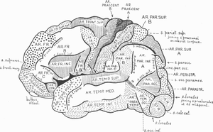
topography of brain cortex
Image via Wikipedia
by Mark Studin DC, FASBE(C), DAAPM, DAAMLP

Chiropractic care improves brain function and the body’s motor or movement ability

Research findings that redefine care for every rehabilitation patient for all motor disorders

According to the Sensory Processing Disorder Foundation (2011), “Sensory processing (sometimes called ‘sensory integration‘ or SI) is a term that refers to the way the nervous system receives messages from the senses and turns them into appropriate motor and behavioral responses. Whether you are biting into a hamburger, riding a bicycle, or reading a book, your successful completion of the activity requires processing sensation or ‘sensory integration'” (http://www.learningrx.com/sensory-motor-integration-faq.htm)

According to Wikipedia (2011), “A motor skill is a learned sequence of movements that combine to produce a smooth, efficient action in order to master a particular task. The development of motor skill occurs in the motor cortex, the region of the cerebral cortex that controls voluntary muscle groups” (http://en.wikipedia.org/wiki/Motor_skill).

According to LearningRX (2010), “Sensory motor integration is the synergistic relationship between the sensory system and the motor system. Since the two communicate and coordinate with each other, if one is problematic, the other can suffer as a result. The two involve receiving and transmitting the stimuli to the central nervous system where the stimulus is then interpreted. The nervous system then determines how to respond and transmits the instructions via nerve impulses to carry out the instructions (e.g. a hand-eye coordination movement)” (http://www.learningrx.com/ sensory-motor-integration-faq.htm).

The synopsis of the above 3 paragraphs is that the human body senses information (sensory processing), processes the information in the brain (sensorimotor cortex), and then sends the information to the part of the body that has to perform a function, such as moving your thumb, walking, talking, picking something up or any other function we do in our lives. As the above paragraph eloquently stated, if any of the 3 areas are not working properly or working not optimally, every part of the system suffers.

In 2010, Taylor and Murphy concluded in their research that chiropractic care improves the functional levels of the motor cortex, premotor areas, and that this improved measurement was maintained after a 20-minute training task, indicating that it wasn’t a transient finding. The authors further offered that the practical applications suggesting that:

1. this alters the way the central nervous system responds to motor training

2. a chiropractic spinal adjustment/manipulation alters the neurological integration at the cortical (brain) level

3. this explains the mechanism responsible for reducing pain levels and increased functional ability after the adjustment/manipulation

4. this explains the mechanism of overuse injuries and chronic pain conditions

The above 4 areas change the way we should approach strategies in rehabilitation for all neurodegenerative and congenital motor and sensory disorders. A list of potential disorders that could benefit in rehabilitation from this research is:

1. muscular dystrophy

2. Duchenne muscular dystrophy

3. myasthenia gravis

4. Parkinson’s disease

5. fibromyalgia

6. multiple sclerosis

7. Huntington’s disease

8. stroke victims

9. all other neuro-muscular diseases

On a clinical note, this author, having cared for muscular dystrophy patients for 30 years, can report that in every instance, the patients were able to ambulate (walk) with greater ease and had significantly more motor control (movement) while under chiropractic care. The goal of rehabilitation in the neurodegenerative patient is to both increase muscle tone and through repetition of activities of daily living, gait training, balance training, speech training and all other motor functions, to help retrain the muscles to maximize the body’s ability to regain those functions. The rehabilitation is essential in most cases and critical to the person regaining an independent life.

The therapist in rehabilitation creates a setting similar to a car or kitchen so that the patient can re-create activities of daily living. In doing these activities with the help of the therapist, the patient is activating stimuli in the sensory nervous system. Touching and movement are senses that the brain has to process and then send impulses back to the muscles to move in order to perform daily tasks. In order for function to be regained maximally, there can be no dysfunction at the spinal level. That dysfunction is defined in chiropractic as subluxation or a vertebrate out of place, negatively affecting the nerve and fixed in the wrong position.

Based upon the research by Taylor and Murphy (2010), if there is a spinal dysfunction (subluxation) it prevents normal impulses from the sensory system and lowers the ability of the brain from functioning at its optimal. Therefore, the most rehabilitation can offer is maximization of the body’s ability at reduced capacity. The implications are staggering as in many cases that could mean no matter the expertise of the therapist or the diligence of the patient, the rehabilitation would not be as successful or could fail if the brain could not function at a higher level.

Through chiropractic care, the patient can have the ability to function at a higher level and live a “more normal life” with neurodegenerative disorders. The implications go well beyond neurodegenerative disorders and cross over to industry, sports and everyday life. However, that will be discussed in another article.

References:

1. Sensory Processing Disorder Foundation (2011). About SPD. Retrieved from http://spdfoundation.net/about-sensory-processing-disorder.html

2. Wikipedia (2011). Motor skill. Retrieved from http://en.wikipedia.org/wiki/Motor_skill

3. LearningRX (2010). Sensory motor integration. Retrieved from http://www.learningrx.com/sensory-motor-integration-faq.htm

4. Taylor, H. H., & Murphy, B. (2010). The effects of spinal manipulation on central integration of dual somatosensory input observed after motor training: A crossover study. Journal of Manipulative and Physiological Therapeutics, 33(4), 261-272.

If you have any further questions. call or contact us at www.nevadachiro.com


Enhanced by Zemanta

Blood Pressure and Your Spine

Main complications of persistent high blood pr...
Image via Wikipedia

Journal of Human Hypertension 2007 (May);   21 (5):   347–352 ~ FULL TEXT

Bakris G, Dickholtz M, Meyer PM, Kravitz G, Avery E, Miller M, Brown J, Woodfield C, Bell B

Department of Preventive Medicine, Rush University Hypertension Center, Chicago, IL, USA

Anatomical abnormalities of the cervical spine at the level of the Atlas vertebra are associated with relative ischaemia of the brainstem circulation and increased blood pressure (BP). Manual correction of this mal-alignment has been associated with reduced arterial pressure. This pilot study tests the hypothesis that correcting mal-alignment of the Atlas vertebra reduces and maintains a lower BP. Using a double blind, placebo-controlled design at a single center, 50 drug naive (n=26) or washed out (n=24) patients with Stage 1 hypertension were randomized to receive a National Upper Cervical Chiropractic (NUCCA) procedure or a sham procedure. Patients received no antihypertensive meds during the 8-week study duration. The primary end point was changed in systolic and diastolic BP comparing baseline and week 8, with a 90% power to detect an 8/5 mm Hg difference at week 8 over the placebo group. The study cohort had a mean age 52.7+/-9.6 years, consisted of 70% males. At week 8, there were differences in systolic BP (-17+/-9 mm Hg, NUCCA versus -3+/-11 mm Hg, placebo; P<0.0001) and diastolic BP (-10+/-11 mm Hg, NUCCA versus -2+/-7 mm Hg; P=0.002). Lateral displacement of Atlas vertebra (1.0, baseline versus 0.04 degrees week 8, NUCCA versus 0.6, baseline versus 0.5 degrees , placebo; P=0.002). Heart rate was not reduced in the NUCCA group (-0.3 beats per minute, NUCCA, versus 0.5 beats per minute, placebo). No adverse effects were recorded. We conclude that restoration of Atlas alignment is associated with marked and sustained reductions in BP similar to the use of two-drug combination therapy.

From the Full-Text Article:

Discussion

The findings of this pilot study represent the first demonstration of a sustained BP lowering effect associated with a procedure to correct the alignment of the Atlas vertebra. The improvement in BP following the correction of Atlas misalignment is similar to that seen by giving two different antihypertensive agents simultaneously. [11, 12] Moreover, this reduction in BP persisted at 8 weeks and was not associated with pain or pain relief or any other symptom that could be associated with a rise in BP.

Other studies support the notion that changes in the cerebral circulation that is related to the position of the Atlas vertebra can affect BP. Coffee et al. [9] reviewed MR images and demonstrated a significant association between pulsatile arterial compression of the ventrolateral medulla and presence of hypertension. They concluded that subjects with hypertension should have an evaluation of their posterior fossa for evidence of anatomic abnormalities. [9 ] In fact, data linking changes in Atlas anatomy and posterior fossa circulatory changes associated with hypertension date back more than 40 years and are reviewed by Reis. [4]

The mechanism as to why this improvement in BP occurs is unknown and cannot be determined by this study. What is clear is that a significant change in sympathetic tone is probably not a major contributing mechanism as heart rate was not significantly changed. The data presented, however, raises a number of important questions including: (a) how does misalignment of C1 affect hypertension? (b) If there is a cause and effect relationship between C1 misalignment and hypertension is malposition of C1 an additional risk factor for the development of hypertension?

What is clear is that misalignment of the Atlas vertebra can be determined by assessment of the alignment of the pelvic crests. This should be considered in those who have a history of hypertension and require multiple medications for treatment. Additionally, it should be considered in those with refractory hypertension and a history of neck injuries, independent of the presence of pain. Note that pain was not present in any of the patients randomized in this study.

As discussed in the Methods section of this paper, techniques are now available to screen for atlas misalignment. This type of screening should be the responsibility of the primary care physician and should be performed on patients who have a history of head and neck trauma even if it is deemed insignificant. Those patients who present with pain related to head and neck trauma should not be screened. At a time when the prevalence of hypertension is increasing and its control more difficult due to a variety of factors, linking the correction of C1 misalignment to the subsequent lowering of BP may represent an important advancement in the screening of such patients.

This pilot study has limitations including the fact that it was dependent on the skill of one practitioner to do the manipulation. It was designed, however, to test the concept that nonsurgical manipulation can alleviate elevations in BP, thus it cannot be generalized until confirmed in a larger trial, which is being planned.

What is known about this topic:

  • The relationship between hypertension and presence of circulatory abnormalities in the area around the Atlas vertebra have been known for more than 40 years [3–5]
  • Recent studies by Akimura et al. [8] noted compression of the ventrolateral branches of the vertebral artery in the area around the Atlas vertebra in 90.6% hypertensive cases
  • Thus, alterations in Atlas anatomy can generate changes in the vertebral circulation that may be associated with elevated levels of blood pressure


What does this study add

  • This pilot study provides evidence that nonsurgical interventions to align the Atlas vertebra provide long-term amelioration in blood pressure
  • It provides the first nonsurgical approach to alleviating elevations in blood pressure


References

1   Randomization of patients Seventh report of the joint national committee on prevention, detection, evaluation, and treatment of high blood pressure
Hypertension 2003;   42:   1206–1252

2   Trends in prevalence, awareness, treatment, and control of hypertension in the United States, 1988–2000
JAMA 2003;   290:   199–206

3 The C1 area of the brainstem in tonic and reflex control of blood pressure. State of the art lecture
Hypertension 1988 (Feb);   11 (2 Pt 2):   18-13

4   The brain and hypertension: reflections on 35 years of inquiry into the neurobiology of the circulation
Circulation 1984;   70:   III31–III45

5   Two specific brainstem systems which regulate the blood pressure
Clin Exp Pharmacol Physiol 1975;   12(Suppl 2):   179–183

6   Neurogenic hypertension: etiology and surgical treatment. I. Observations in 53 patients
Ann Surg 1985;   201:   391–398

7   Microvascular decompression in the treatment of hypertension: review and update
Surg Neurol 2001;   55:   2–10

8   Essential hypertension and neurovascular compression at the ventrolateral medulla oblongata: MR evaluation
AJNR Am J Neuroradiol 1995 (Feb);   16 (2):   401-5

9   Arterial compression of the retro-olivary sulcus of the medulla in essential hypertension: a multivariate analysis
J Hypertens 2005 (Nov);   23 (11):   2027-31

10   Arterial compression of the retro-olivary sulcus of the ventrolateral medulla in essential hypertension and diabetes
Hypertension 2005;   46:   982–985

11   Combination drug treatment for hypertension with nondiabetic renal disease
Curr Hypertens Rep 2005;   7:   358–359

12   Achieving goal blood pressure in patients with type 2 diabetes: conventional versus fixed-dose combination approaches
J Clin Hypertens 2003;   5:   202–209

Enhanced by Zemanta

What You Need to Know About Belly Fat

Excess adipose tissue around a male's mid-section.
Image via Wikipedia

The abdominal region has two types of fat: 1. Subcutaneous fat, which lies directly beneath the skin and on top of the abdominal muscles, and 2. Visceral fat, which is deeper in the abdomen beneath your muscle and surrounds the organs. Visceral fat is what gives men their “beer belly”. This beer belly looks soft but is quite hard to touch. This is an enormous storage area of toxins and indicates blood sugar problems.

Subcutaneous fat and visceral fat are a health risk, but studies have shown the excessive visceral fat is even more dangerous than subcutaneous fat. Both types of excessive fats increase the risk of developing heart disease, diabetes, high blood pressure, stroke, sleep apnea, various forms of cancer, and other degenerative diseases.

The reason visceral fat is the most dangerous is that it releases more inflammatory molecules into your body on a consistent basis. Bad fats create more bad fats and other toxins. Because the body cannot eliminate the toxins via the liver and kidneys, the body puts the toxins back into the blood stream, which will surround toxins with fluid and fat to dilute and insulate the poisonous effect.

The ONLY solution to consistently lose your abdominal fat and keep it off for good is to combine a sound nutritious diet full of unprocessed natural foods with a specific nutritional supplementation program, designed for that person’s individual needs. Nutritious foods and proper individualized supplementation needs to be combined with a consistent exercise program that stimulates the necessary hormonal and metabolic response within your body. Your food and supplement intake, as well as your training program is important for your looks and your health.

We have the key to unlocking the body’s ability to properly and efficiently clean up the damage and nutrient the body for permanent and lasting weight loss.

For more information, go to www.nevadachiro.com or call 565-7474.

Enhanced by Zemanta

Forward Head Posture and TMJ

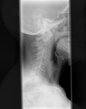
Xray of cervical spine
Image via Wikipedia

Forward head posture creates a domino effect in the body:

  • Moving forward, the head shifts the center of gravity.
  • To compensate, the upper body drifts backward.
  • To compensate for the upper body shift, the hips tilt forward.

So, the forward head position can be the cause of not only the head, neck and TMJ problems, but also mid back and low back problems. We can gain a better understanding of the interaction between the mouth opening and closing, TMJ bio-mechanics, and neck  function with an overview of the structures involved.

According to Rene Cailliet, Physical Medicine and Rehabilitation Director at U.S.C. states: “It’s an axiom … that the body follows the head … You can realign your entire body by moving your head … your head held in a forward position can pull your entire body out of line.” He goes on to explain that the vital lung capacity is reduced as much as 30%. The gastrointestinal system is affected, particularly the large intestine. When a hunched position is assumed, the body becomes rigid, and range of motion is affected. Since endorphin production is reduced, an increase in pain and discomfort results.

Another researcher Kapandji, in his classic text on spinal function states, “The anterior muscles of the neck … act as the long arm of a lever … they are powerful flexors of the head and cervical column … flattening the cervical column.”

Additionally, numerous investigators describe the effect of altered jaw position on head posture. Forward and side to side head position changes the jaw, throat, and tongue. It compresses the neck  joints causing muscular nerve entrapments. Nerve root compression or posterior neck vertebral joint irritation or restriction result in peripheral entrapment neuropathies. One common entrapment is the greater or lesser suboccipital nerves that pass between the occiput and atlas. This may cause headaches or refer pain to the facial region.

General Symptoms by Head Forward Posture problems
A. Chronically tired (lowered hemoglobin, immature cells) 89%
B. Increased nervous tension 86%
C. Malaise 61%
D. Restless sleep (awaken tired) 78%
E. Numbness in hands (awaken with arm or leg asleep) 32%
F. Cold hands and feet (poor circulation) 67%
G. Back aches and leg aches (tired, ache-all-over feeling) 47%
H. Thirsty (much water doesn’t satisfy) 43%
I. Restless nibbling at food (never satisfied)
J. Blood (variations in count and quality; many irregularly formed cells; cell walls even thickness one week after treatment)
K. Lowered hemoglobin
L. Lower thyroid activity (an almost constant finding)
M. Facial pallor
N. Dull, non-sparkling eyes

Forward head posture is an enormous problem of our society. Nearly 100% of new patients presenting in my office have it. If you have it and don’t correct it, it will lead to neck arthritis and damaged discs.

If you are interested in being checked for forward head posture or want more information visit www.nevadachiro.com.

Enhanced by Zemanta