The Facts about Gastroesophageal Reflux Disorder

I wanted to share this article by one of my mentors, Dr. Howard Loomis. He is the godfather of enzyme replacement therapy.

by Howard Loomis, D.C.

Before beginning this discussion, I wish to point out that current diagnosis of this seemingly pandemic condition is done without physical examination or objective testing. Recommendation of antacids, proton-pump inhibitors, and histamine antagonists, thus canceling normal digestion in the stomach, can only lead to chronic degenerative conditions.

GERD is a condition that is described as occurring when the acidified liquid contents of the stomach back up into the esophagus. This includes not only stomach acid, but pepsin as well and may even include bile that has backed-up into the stomach from the duodenum. The liquid can inflame and damage the lining of the esophagus, if it is not protected by the mucosal lining.  This is in and of itself not a disease.

The symptoms of uncomplicated GERD are heartburn, regurgitation, and nausea, but these symptoms only occur in a minority of patients. This statement requires some explanation.

Reflux of the stomach’s liquid contents into the esophagus occurs in most normal individuals. In fact, one study found that reflux occurs as frequently in normal individuals as in patients with a diagnosis of GERD. There are several mechanisms that prevent the symptoms from occurring:

  • Most reflux occurs during the day when we are upright and refluxed liquid is more likely to flow back down into the stomach due to the effect of gravity.
  • When awake we repeatedly swallow and each swallow carries any refluxed liquid back into the stomach. Also saliva contains bicarbonate and with each swallow it travels down the esophagus and neutralizes the small amount of acid that remains in the esophagus after gravity and swallowing have removed most of the liquid.
  • At night while sleeping, gravity is not in effect, swallowing stops, and the secretion of saliva is reduced. Therefore, reflux that occurs at night is more likely to result in acid remaining in the esophagus longer and can cause damage to the esophagus.

Symptoms of Uncomplicated GERD

Heartburn
When acid refluxes back into the esophagus in patients with GERD, nerve fibers in the esophagus are stimulated. This nerve stimulation results most commonly in heartburn, the pain that is characteristic of GERD. Heartburn usually is described as a burning pain in the middle of the chest. It may start high in the abdomen or may extend up into the neck. In some patients, however, the pain may be sharp or pressure-like, rather than burning. Such pain can mimic heart pain or angina. In other patients, the pain may extend to the back.

Episodes of heartburn tend to happen periodically. This means that the episodes are more frequent or severe for a period of several weeks or months, and then they become less frequent or severe or even absent for several weeks or months. Nevertheless, medicine considers GERD to be a chronic condition and once treatment for GERD is begun it is continued indefinitely.

Regurgitation
Usually only small quantities of liquid reach the esophagus, and the liquid remains in the lower esophagus. At the upper end of the esophagus is the upper esophageal sphincter (UES). The upper esophageal sphincter is a circular ring of muscle that is very similar in its actions to the lower esophageal sphincter (LES). That is, it prevents esophageal contents from backing up into the throat. When small amounts of refluxed liquid and/or foods pass back through the UES and enter the throat, there may be an acid taste in the mouth. If larger quantities breach the UES, patients may suddenly find their mouths filled with the liquid or food.

Nausea
Nausea is uncommon in GERD. In some patients, however, it may be frequent or severe and may result in vomiting. In fact, in patients with unexplained nausea and/or vomiting, GERD is one of the first conditions suspected. It is not clear why some patients with GERD develop mainly heartburn and others develop mainly nausea.

The Causes of GERD

These include an abnormal lower esophageal sphincter, weak or abnormal esophageal contractions, hiatal hernia, and slow emptying of the stomach – BUT NOT EXCESS STOMACH ACID PRODUCTION, except in the rare Zollinger-Ellison Syndrome. A small number of patients with GERD do produce abnormally large amounts of acid, but this is uncommon and not a contributing factor in the vast majority of patients.

Lower esophageal sphincter (C3 to C5 and T4 to T9)
The action of the lower esophageal sphincter (LES) is the most important factor for preventing reflux. The LES is a specialized ring of muscle that surrounds the lower-most end of the esophagus where it joins the stomach. This muscle is active most of the time, contracting and closing off the passage from the esophagus into the stomach. When food or saliva is swallowed, the LES relaxes for a few seconds to allow the food or saliva to pass into the stomach, and then it closes again. Two abnormalities of the LES have been found in patients with GERD:

  • The first is abnormally weak contraction of the LES, which reduces its ability to prevent reflux.
  • The second is abnormal relaxations of the LES that do not accompany swallows and they can last up to several minutes.

These prolonged relaxations allow reflux to occur more easily. The transient LES relaxations occur in patients with GERD most commonly after meals when the stomach is distended with food.

Hiatal hernia
Hiatal hernias may contribute to reflux, but not necessarily. A majority of patients with GERD have hiatal hernias, but many do not AND many people have hiatal hernias but do not have GERD! There appear to be three reasons why a hiatal hernia can contribute to GERD:

  • First, the LES is located at the same level where the esophagus passes from the chest through the diaphragm and into the abdomen. When there is a hiatal hernia, a small part of the upper stomach that attaches to the esophagus pushes up through the diaphragm. As a result, a small part of the stomach and the LES come to lie in the chest, and the LES is no longer at the level of the diaphragm. This means the barrier to reflux is equal to the sum of the pressures generated by the LES and the diaphragm.
  • Second, a hiatal hernia produces a small pouch of stomach above the diaphragm. The sac is pinched off from the esophagus above by the LES and from the stomach below by the diaphragm. The sac traps acid that comes from the stomach and it is easier for the acid to reflux when the LES relaxes with a swallow or a transient relaxation.
  • Finally, the esophagus normally joins the stomach obliquely, not straight on or at a 90-degree angle. Due to this oblique angle of entry, a flap of tissue is formed between the stomach and esophagus. This flap acts like a valve, shutting off the esophagus from the stomach and preventing reflux. When a hiatal hernia allows entry of the esophagus into the stomach the valve-like flap is distorted or disappears and it no longer can help prevent reflux.

Esophageal contractions
Swallowing is important in eliminating acid in the esophagus. Swallowing causes a ring-like wave of contraction of the esophageal muscles, which narrows the lumen of the esophagus. Peristalsis begins in the upper esophagus and travels to the lower esophagus. It pushes food, saliva, and whatever else is in the esophagus into the stomach. If the contraction is weak, refluxed acid cannot be pushed back into the stomach.

Abnormalities of contraction are found frequently in patients with GERD. In fact, they are found most frequently in those patients with the most severe GERD. Note that smoking also substantially reduces the clearance of acid from the esophagus. This effect continues for at least 6 hours after the last cigarette.

Delayed Emptying of the stomach
A minority of patients with GERD, about 20%, have been found to have stomachs that empty more slowly than normal after a meal. This slower emptying of the stomach prolongs the distention of the stomach with food after meals. Therefore, the slower emptying prolongs the period of time during which reflux is more likely to occur.

Hi. Dr. Swolensky again! If you have questions about this information please contact my office at www.nevadachiro.com or call (702) 565-7474.

Enhanced by Zemanta

Brain Function (Sensorimotor Cortex) Increases with Chiropractic Care

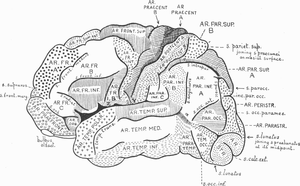
topography of brain cortex
Image via Wikipedia
by Mark Studin DC, FASBE(C), DAAPM, DAAMLP

Chiropractic care improves brain function and the body’s motor or movement ability

Research findings that redefine care for every rehabilitation patient for all motor disorders

According to the Sensory Processing Disorder Foundation (2011), “Sensory processing (sometimes called ‘sensory integration‘ or SI) is a term that refers to the way the nervous system receives messages from the senses and turns them into appropriate motor and behavioral responses. Whether you are biting into a hamburger, riding a bicycle, or reading a book, your successful completion of the activity requires processing sensation or ‘sensory integration'” (http://www.learningrx.com/sensory-motor-integration-faq.htm)

According to Wikipedia (2011), “A motor skill is a learned sequence of movements that combine to produce a smooth, efficient action in order to master a particular task. The development of motor skill occurs in the motor cortex, the region of the cerebral cortex that controls voluntary muscle groups” (http://en.wikipedia.org/wiki/Motor_skill).

According to LearningRX (2010), “Sensory motor integration is the synergistic relationship between the sensory system and the motor system. Since the two communicate and coordinate with each other, if one is problematic, the other can suffer as a result. The two involve receiving and transmitting the stimuli to the central nervous system where the stimulus is then interpreted. The nervous system then determines how to respond and transmits the instructions via nerve impulses to carry out the instructions (e.g. a hand-eye coordination movement)” (http://www.learningrx.com/ sensory-motor-integration-faq.htm).

The synopsis of the above 3 paragraphs is that the human body senses information (sensory processing), processes the information in the brain (sensorimotor cortex), and then sends the information to the part of the body that has to perform a function, such as moving your thumb, walking, talking, picking something up or any other function we do in our lives. As the above paragraph eloquently stated, if any of the 3 areas are not working properly or working not optimally, every part of the system suffers.

In 2010, Taylor and Murphy concluded in their research that chiropractic care improves the functional levels of the motor cortex, premotor areas, and that this improved measurement was maintained after a 20-minute training task, indicating that it wasn’t a transient finding. The authors further offered that the practical applications suggesting that:

1. this alters the way the central nervous system responds to motor training

2. a chiropractic spinal adjustment/manipulation alters the neurological integration at the cortical (brain) level

3. this explains the mechanism responsible for reducing pain levels and increased functional ability after the adjustment/manipulation

4. this explains the mechanism of overuse injuries and chronic pain conditions

The above 4 areas change the way we should approach strategies in rehabilitation for all neurodegenerative and congenital motor and sensory disorders. A list of potential disorders that could benefit in rehabilitation from this research is:

1. muscular dystrophy

2. Duchenne muscular dystrophy

3. myasthenia gravis

4. Parkinson’s disease

5. fibromyalgia

6. multiple sclerosis

7. Huntington’s disease

8. stroke victims

9. all other neuro-muscular diseases

On a clinical note, this author, having cared for muscular dystrophy patients for 30 years, can report that in every instance, the patients were able to ambulate (walk) with greater ease and had significantly more motor control (movement) while under chiropractic care. The goal of rehabilitation in the neurodegenerative patient is to both increase muscle tone and through repetition of activities of daily living, gait training, balance training, speech training and all other motor functions, to help retrain the muscles to maximize the body’s ability to regain those functions. The rehabilitation is essential in most cases and critical to the person regaining an independent life.

The therapist in rehabilitation creates a setting similar to a car or kitchen so that the patient can re-create activities of daily living. In doing these activities with the help of the therapist, the patient is activating stimuli in the sensory nervous system. Touching and movement are senses that the brain has to process and then send impulses back to the muscles to move in order to perform daily tasks. In order for function to be regained maximally, there can be no dysfunction at the spinal level. That dysfunction is defined in chiropractic as subluxation or a vertebrate out of place, negatively affecting the nerve and fixed in the wrong position.

Based upon the research by Taylor and Murphy (2010), if there is a spinal dysfunction (subluxation) it prevents normal impulses from the sensory system and lowers the ability of the brain from functioning at its optimal. Therefore, the most rehabilitation can offer is maximization of the body’s ability at reduced capacity. The implications are staggering as in many cases that could mean no matter the expertise of the therapist or the diligence of the patient, the rehabilitation would not be as successful or could fail if the brain could not function at a higher level.

Through chiropractic care, the patient can have the ability to function at a higher level and live a “more normal life” with neurodegenerative disorders. The implications go well beyond neurodegenerative disorders and cross over to industry, sports and everyday life. However, that will be discussed in another article.

References:

1. Sensory Processing Disorder Foundation (2011). About SPD. Retrieved from http://spdfoundation.net/about-sensory-processing-disorder.html

2. Wikipedia (2011). Motor skill. Retrieved from http://en.wikipedia.org/wiki/Motor_skill

3. LearningRX (2010). Sensory motor integration. Retrieved from http://www.learningrx.com/sensory-motor-integration-faq.htm

4. Taylor, H. H., & Murphy, B. (2010). The effects of spinal manipulation on central integration of dual somatosensory input observed after motor training: A crossover study. Journal of Manipulative and Physiological Therapeutics, 33(4), 261-272.

If you have any further questions. call or contact us at www.nevadachiro.com


Enhanced by Zemanta

Beyond Antibiotics: Are There Healthier Options?

by Dr. Michael Schmidt

For decades, antibiotics have been the cornerstone of treatment for bacterial infections. Antibiotics have saved many lives during this time. However, doctors have come to rely too heavily on these miracle drugs and the overuse of antibiotics has led to serious problems.

For all of the potential benefits of antibiotics, a growing list of adverse health consequences has emerged because of overuse. A sample of these is listed below:

  • Children with chronic earaches who take many series of antibiotics experience 2-6 times more recurrent ear infections.
  • Antibiotic overuse can upset intestinal integrity.
  • Antibiotic overuse can suppress immune function.

Factors that Lower Immunity . We know that there are many factors that lower immunity and thus make the body more susceptible to infection. These include:

  • Nutrition: Low vitamin C and zinc lead to sluggish Immune response.
  • Diet: Excess sugar consumption slows the ability of white blood cells to engulf and destroy bacteria.
  • Lifestyle: Sedentary people tend to have more sluggish immune systems than active people.
  • Environment: Solvent chemicals and heavy metals such as lead increase susceptibility to infection.
  • Psychological: People under stress are more likely to have frequent infections, while those with better coping skills are less sensitive to the effects of stress on their immune systems.

Are there Natural Remedies that can Help? For many common ailments, scientific research is confirming time-honored traditions.

  • Ear infections: The vast majority of children with recurrent ear infections improved after removing food allergens from their diets. Warm sesame oil drops can help ease the pain.
  • Bladder infections: Unsweetened cranberry juice clears bacteria from the bladder. Yogurt and acidophilus are also helpful.
  • Respiratory infections: A sauna once a week can reduce respiratory problems by one- half.
  • Intestinal infections: Homeopathic medicine has been helpful in reducing diarrhea in children.

Live Healthfully and Be Informed. Remember, antibiotics can save lives and reduce suffering. They are vital components of the healing arsenal. However, they must be used wisely. Your goal should be to optimize the health of your family to reduce the need for drug intervention and to make wise, informed decisions should drug intervention become necessary.

Are Antibiotics Being Overused in Your Care? Does your doctor:

  • Prescribe antibiotics over the phone?
  • Refill a prescription without an examination?
  • Prescribe without taking a blood count?
  • Neglect to ask about diet, nutrition or lifestyle factors?- Prescribe after only a cursory examination?
  • Write off or ignore concerns about negative side effects?
  • Remark that antibiotics are harmless?
  • Try to intimidate or frighten you into following orders?
  • Prescribe several courses of antibiotics when there is no improvement?
  • Prescribe antibiotics when illness is viral such as a cold?
  • Seem overworked or overbooked?
  • Cut your visit short by handing you a prescription?

If you answered “yes” to more than five items, you or someone in your family may be receiving antibiotics needlessly. Ask your doctor for a full re-evaluation or seek another opinion.

Dr. Schmidt‘s books Beyond Antibiotics: Strategies for Living in a World of Emerging Infections & Antibiotic-Resistant Bacteria -Third Edition and Healing Childhood Ear Infections: Prevention, Home Care, and Alternative Treatment . Both can both be ordered through DDR‘s Amazon Associates account which provides financial support to the organization.

If you would like more information call 565-7474. Or go to our website: www.nevadachiro.com

Enhanced by Zemanta

Blood Pressure and Your Spine

Main complications of persistent high blood pr...
Image via Wikipedia

Journal of Human Hypertension 2007 (May);   21 (5):   347–352 ~ FULL TEXT

Bakris G, Dickholtz M, Meyer PM, Kravitz G, Avery E, Miller M, Brown J, Woodfield C, Bell B

Department of Preventive Medicine, Rush University Hypertension Center, Chicago, IL, USA

Anatomical abnormalities of the cervical spine at the level of the Atlas vertebra are associated with relative ischaemia of the brainstem circulation and increased blood pressure (BP). Manual correction of this mal-alignment has been associated with reduced arterial pressure. This pilot study tests the hypothesis that correcting mal-alignment of the Atlas vertebra reduces and maintains a lower BP. Using a double blind, placebo-controlled design at a single center, 50 drug naive (n=26) or washed out (n=24) patients with Stage 1 hypertension were randomized to receive a National Upper Cervical Chiropractic (NUCCA) procedure or a sham procedure. Patients received no antihypertensive meds during the 8-week study duration. The primary end point was changed in systolic and diastolic BP comparing baseline and week 8, with a 90% power to detect an 8/5 mm Hg difference at week 8 over the placebo group. The study cohort had a mean age 52.7+/-9.6 years, consisted of 70% males. At week 8, there were differences in systolic BP (-17+/-9 mm Hg, NUCCA versus -3+/-11 mm Hg, placebo; P<0.0001) and diastolic BP (-10+/-11 mm Hg, NUCCA versus -2+/-7 mm Hg; P=0.002). Lateral displacement of Atlas vertebra (1.0, baseline versus 0.04 degrees week 8, NUCCA versus 0.6, baseline versus 0.5 degrees , placebo; P=0.002). Heart rate was not reduced in the NUCCA group (-0.3 beats per minute, NUCCA, versus 0.5 beats per minute, placebo). No adverse effects were recorded. We conclude that restoration of Atlas alignment is associated with marked and sustained reductions in BP similar to the use of two-drug combination therapy.

From the Full-Text Article:

Discussion

The findings of this pilot study represent the first demonstration of a sustained BP lowering effect associated with a procedure to correct the alignment of the Atlas vertebra. The improvement in BP following the correction of Atlas misalignment is similar to that seen by giving two different antihypertensive agents simultaneously. [11, 12] Moreover, this reduction in BP persisted at 8 weeks and was not associated with pain or pain relief or any other symptom that could be associated with a rise in BP.

Other studies support the notion that changes in the cerebral circulation that is related to the position of the Atlas vertebra can affect BP. Coffee et al. [9] reviewed MR images and demonstrated a significant association between pulsatile arterial compression of the ventrolateral medulla and presence of hypertension. They concluded that subjects with hypertension should have an evaluation of their posterior fossa for evidence of anatomic abnormalities. [9 ] In fact, data linking changes in Atlas anatomy and posterior fossa circulatory changes associated with hypertension date back more than 40 years and are reviewed by Reis. [4]

The mechanism as to why this improvement in BP occurs is unknown and cannot be determined by this study. What is clear is that a significant change in sympathetic tone is probably not a major contributing mechanism as heart rate was not significantly changed. The data presented, however, raises a number of important questions including: (a) how does misalignment of C1 affect hypertension? (b) If there is a cause and effect relationship between C1 misalignment and hypertension is malposition of C1 an additional risk factor for the development of hypertension?

What is clear is that misalignment of the Atlas vertebra can be determined by assessment of the alignment of the pelvic crests. This should be considered in those who have a history of hypertension and require multiple medications for treatment. Additionally, it should be considered in those with refractory hypertension and a history of neck injuries, independent of the presence of pain. Note that pain was not present in any of the patients randomized in this study.

As discussed in the Methods section of this paper, techniques are now available to screen for atlas misalignment. This type of screening should be the responsibility of the primary care physician and should be performed on patients who have a history of head and neck trauma even if it is deemed insignificant. Those patients who present with pain related to head and neck trauma should not be screened. At a time when the prevalence of hypertension is increasing and its control more difficult due to a variety of factors, linking the correction of C1 misalignment to the subsequent lowering of BP may represent an important advancement in the screening of such patients.

This pilot study has limitations including the fact that it was dependent on the skill of one practitioner to do the manipulation. It was designed, however, to test the concept that nonsurgical manipulation can alleviate elevations in BP, thus it cannot be generalized until confirmed in a larger trial, which is being planned.

What is known about this topic:

  • The relationship between hypertension and presence of circulatory abnormalities in the area around the Atlas vertebra have been known for more than 40 years [3–5]
  • Recent studies by Akimura et al. [8] noted compression of the ventrolateral branches of the vertebral artery in the area around the Atlas vertebra in 90.6% hypertensive cases
  • Thus, alterations in Atlas anatomy can generate changes in the vertebral circulation that may be associated with elevated levels of blood pressure


What does this study add

  • This pilot study provides evidence that nonsurgical interventions to align the Atlas vertebra provide long-term amelioration in blood pressure
  • It provides the first nonsurgical approach to alleviating elevations in blood pressure


References

1   Randomization of patients Seventh report of the joint national committee on prevention, detection, evaluation, and treatment of high blood pressure
Hypertension 2003;   42:   1206–1252

2   Trends in prevalence, awareness, treatment, and control of hypertension in the United States, 1988–2000
JAMA 2003;   290:   199–206

3 The C1 area of the brainstem in tonic and reflex control of blood pressure. State of the art lecture
Hypertension 1988 (Feb);   11 (2 Pt 2):   18-13

4   The brain and hypertension: reflections on 35 years of inquiry into the neurobiology of the circulation
Circulation 1984;   70:   III31–III45

5   Two specific brainstem systems which regulate the blood pressure
Clin Exp Pharmacol Physiol 1975;   12(Suppl 2):   179–183

6   Neurogenic hypertension: etiology and surgical treatment. I. Observations in 53 patients
Ann Surg 1985;   201:   391–398

7   Microvascular decompression in the treatment of hypertension: review and update
Surg Neurol 2001;   55:   2–10

8   Essential hypertension and neurovascular compression at the ventrolateral medulla oblongata: MR evaluation
AJNR Am J Neuroradiol 1995 (Feb);   16 (2):   401-5

9   Arterial compression of the retro-olivary sulcus of the medulla in essential hypertension: a multivariate analysis
J Hypertens 2005 (Nov);   23 (11):   2027-31

10   Arterial compression of the retro-olivary sulcus of the ventrolateral medulla in essential hypertension and diabetes
Hypertension 2005;   46:   982–985

11   Combination drug treatment for hypertension with nondiabetic renal disease
Curr Hypertens Rep 2005;   7:   358–359

12   Achieving goal blood pressure in patients with type 2 diabetes: conventional versus fixed-dose combination approaches
J Clin Hypertens 2003;   5:   202–209

Enhanced by Zemanta

Why You NEED to Care About Your Spine

Центральная нервная система-Central nervous system
Image via Wikipedia

The following is deep and steeped with big words. However, the words also include definitions, so don’t let it stop you from reading. It’s written by a mentor of mine and I wanted you to get a much fuller and deeper understanding of what chiropractic is and why you NEED to know!

If you have any questions go to www.nevadachiro.com or call 565-7474.

A Four-Dimensional Model of Vertebral Subluxation

By Christopher Kent, DC, Esq.

Vertebral subluxation represents the heart and soul of chiropractic, yet to many chiropractors, it remains a clinical conundrum. I believe that the controversy and confusion surrounding the chiropractic concept of vertebral subluxation is due, in part, to the lack of an operational definition compatible with most techniques.

A review of models of vertebral subluxation has been published elsewhere.1 However, regardless of the elegance of a theoretical model, it must be capable of being operationalized if it is to be used to develop clinical strategies. The four-dimensional model was developed as an initial step in the operational definition of vertebral subluxation. It incorporates traditional chiropractic constructs and serves as a bridge to contemporary technology.

The First Dimension

The traditional safety-pin cycle (SPC) consists of the transmission of afferent information from the tissue cell (periphery) to the brain cell (epiphery) on one side, and on the efferent side, from brain cell to tissue cell.2 In the 4-D model, the first dimension is dysafferentation, representing the afferent portion of the SPC. Aberrant afferent input to the CNS may result in qualitatively and/or quantitatively inappropriate responses to changes in the internal or external environment.1 In the contemporary jargon of the computer industry, there is “garbage in – garbage out.” Dr. Fred Barge, in his book One Cause, One Cure, stated that the cause of disease is “The body’s inability to comprehend itself and/or its environment.”3 Such “comprehension” is dependent upon interference-free afferent input.

Dye4 quoted remarks, attributed to B.J. Palmer in August 1935, which express this concept, noting the result of an adjustment: “[T]he restoration of the normal transmission of mental impulse supply from its point of origin within the brain to its point of expression in the diseased part of the body, or vice versa, that the Innate Intelligence within the brain may receive correct, accurate, exact messages as to the external conditions existing at the periphery so that it may direct either the necessary reparative forces or the necessary cooperative forces from that the tissues may be repaired or that the organ or structure may be properly directed that it may perform the normal functioning desired and indicated by the incoming message from me part without.”

The authors of the remarkable book Segmental Neuropathy, published by Canadian Memorial Chiropractic College, proposed the concept of a “neural image,” dependent upon the integrity of neural receptors and afferent pathways. This “neural image” is a representation of the organism’s perception of the external and internal environment. If afferent input is compromised, efferent response may be qualitatively and quantitatively compromised.5

The clinical implications of aberrant or suboptimal afferent go beyond short-term homeostatic regulation. Dysafferentation may result in anatomical and functional changes in the brain itself. Merzenich6 noted, “The brain was constructed to change.” This challenge to the conventional world view that the mature adult brain is stable and unchanging, the only exception being the death of brain cells, has profound implications for the chiropractor.

Gage7 stated, “Researchers first demonstrated that the central nervous systems of mammals contain some innate regenerative properties in the 1960s and 1970s, when several groups showed that axons, or main branches, of neurons in the adult brain and spinal cord can regrow to some extent after injury.” The ability of the brain to change both anatomically and functionally is known as neuroplasticity. Three types have been described:8

  • Experience-independent plasticity refers to changes which are not the result of environmental changes or influence.
  • Experience-expectant plasticity occurs when the brain uses input from the external environment to effect normal developmental changes in its structure.
  • Experience-dependent plasticity is when a modification to the internal or external environment produces change in a feature of the brain.

Holloway6 explained how the brain reconfigures itself and the implications of doing so: “Change the input, be it a behavior, a mental exercise … or a physical skill and the brain changes accordingly. Magnetic resonance imaging machines reveal the new map: different regions light up … [T]he brain can be extensively remodeled throughout the course of one’s life, without drugs, without surgery. Regions of the brain can be taught to do different tasks if need be … This sort of thing will be a part of normal future life … healing plasticity can be driven by behavior.”

The 2nd-4th Dimensions

The remaining three dimensions represent the efferent portion of the safety-pin cycle. Each component may be reliably measured. These measurements provide objective evidence concerning manifestations of vertebral subluxation. The three components are:

  • Dyskinesia. Dyskinesia refers to distortion or impairment of voluntary movement.9 Spinal motion may be reliably measured using inclinometry.10 Alterations in regional ranges of motion are associated with vertebral subluxation.11
  • Dysponesis. Dysponesis is abnormal involuntary muscle activity. Dysponesis refers to a reversible physiopathologic state, consisting of errors in energy expenditure which are capable of producing functional disorders. Dysponesis consists mainly of covert errors in action potential output from the motor and premotor areas of the cortex and the consequences of that output. These neurophysiological reactions may result from responses to environmental events, bodily sensations, and emotions. The resulting aberrant muscle activity may be evaluated using surface electrode techniques.12 Typically, static surface electromyography (SMEG) with axial loading is used to evaluate innate responses to gravitational stress.13
  • Dysautronomia. The autonomic nervous system regulates the actions of organs, glands, and blood vessels. Acquired dysautonomia may be associated with a broad array of functional abnormalities.14-19 Autonomic dystonia may be evaluated by measuring skin temperature differentials.20 Uematsu, et al., determined normative values for skin temperature differences based upon asymptomatic “normal” individuals. The authors stated, “These values can be used as a standard in assessment of sympathetic nerve function, and the degree of asymmetry is a quantifiable indicator of dysfunction … Deviations from the normal values will allow suspicion of neurological pathology to be quantitated and therefore can improve assessment and lead to proper clinical management.”21 Skin temperature differentials are associated with vertebral subluxation.22 Autonomic tone and balance may also be evaluated by measuring heart rate variability.23

This four-dimensional model may be used with any technique which has, as its objective, the detection, management or correction of vertebral subluxation. Correction or reduction of vertebral subluxation facilitates the restoration of proper tone throughout the nervous system. Alterations in the tone of the somatic system may be objectively evaluated using surface EMG. Altered autonomic tone may be evaluated using skin temperature measurements. Changes in ranges of motion may be measured to assess dyskinesia. Such objective assessments have the potential to make chiropractic the dominant strategy of 21st century health care.

References

  1. Kent C. Models of vertebral subluxation: a review. Journal of Vertebral Subluxation Research, 1996;1(1):11.
  2. Stephenson RW. Chiropractic Textbook. The Palmer School of Chiropractic: Davenport, IA. 1948 edition, page 9.
  3. Barge FH. One Cause, One Cure. LaCrosse, WI, 1990.
  4. Dye AA. The Evolution of Chiropractic. Published by A.A. Dye, DC: Philadelphia, 1939, page 266.
  5. Segmental Neuropathy. Canadian Memorial Chiropractic College. Toronto, Ontario. No date. Presumed to be written in the 1960s primarily by H.M. Himes and A. Peterson.
  6. Holloway M. “The Mutable Brain.” Scientific American, 2002;289(3):79.
  7. Gage FH. “Brain, Repair Yourself.” Scientific American, 2002;289(3):47.Neuroplasticity. PowerPoint lecture. www.snl.salk.edu/~nikoosh/Lecture_5.ppt
  8. Dorland’s Pocket Medical Dictionary., 25th Edition. WB Saunders Company, 1995.
  9. Saur PM, Ensink FB, Frese K, et al. Lumbar range of motion: reliability and validity of the inclinometer technique in the clinical measurement of trunk flexibility. Spine, 1996;21(11):1332.
  10. Blunt KL, Gatterman MI, Bereznick DE. Kinesiology: An Essential Approach Toward Understanding the Chiropractic Subluxation. Chapter 11 in Gatterman MI (ed): Foundations of Chiropractic Subluxation. Mosby: St. Louis, MO, 1995.
  11. Whatmore GB, Kohi DR. Dysponesis: a neurophysiologic factor in functional disorders. Behav Sci, 1968;13(2):102.
  12. Kent C. Surface electromyography in the assessment of changes in paraspinal muscle activity associated with vertebral subluxation: a review. Journal of Vertebral Subluxation Research, 1997;1(3):15.
  13. Backonja M-M. Reflex sympathetic dystrophy/sympathetically mediated pain/causalgia: the syndrome of neuropathic pain with dysautonomia. Seminars in Neurology, 1994;14(3):263.
  14. Goldstein DS, Holmes C, Cannon III RO, et al. Sympathetic cardioneuropathy in dysautonomias. New Engl J Med, 1997;336(10):696.
  15. Vassallo M, Camilleri M, Caron BL, Low PA. Gastrointestinal motor dysfunction in acquired selective cholinergic dysautonomia associated with infectious mononucleosis. Gastroenterology, 1991;100(1):252.
  16. Baron R, Engler F. Postganglionic cholinergic dysautonomia with incomplete recovery: a clinical, neurophysiological and immunological case study. J Neurol, 1996;243:18.
  17. Soares JLD. Disautonomias. Acta Medica Portuguesa, 1995;8(7- 8):425. Written in Portuguese. English abstract.
  18. Stryes KS. The phenomenon of dysautonomia and mitral valve prolapse. J Am Acad Nurse Practitioners, 1994;6(1):11.
  19. Korr IM. The Collected Papers of Irvin M. Korr. American Academy of Osteopathy: Indianapolis, IN, 1979.
  20. Uematsu S, Edwin DH, Jankel ER, et al. Quantification of thermal asymmetry. J Neurosurg, 1988;69:552.
  21. Kent C, Gentempo P. “Instrumentation and Imaging in Chiropractic: A Centennial Retrospective.” Today’s Chiropractic, 1995;24(1):32.
  22. Heart rate variability. Standards of measurement, physiological interpretation, and clinical use. Circulation, 1996;93:1043-1065.
Enhanced by Zemanta

What You Need to Know About Belly Fat

Excess adipose tissue around a male's mid-section.
Image via Wikipedia

The abdominal region has two types of fat: 1. Subcutaneous fat, which lies directly beneath the skin and on top of the abdominal muscles, and 2. Visceral fat, which is deeper in the abdomen beneath your muscle and surrounds the organs. Visceral fat is what gives men their “beer belly”. This beer belly looks soft but is quite hard to touch. This is an enormous storage area of toxins and indicates blood sugar problems.

Subcutaneous fat and visceral fat are a health risk, but studies have shown the excessive visceral fat is even more dangerous than subcutaneous fat. Both types of excessive fats increase the risk of developing heart disease, diabetes, high blood pressure, stroke, sleep apnea, various forms of cancer, and other degenerative diseases.

The reason visceral fat is the most dangerous is that it releases more inflammatory molecules into your body on a consistent basis. Bad fats create more bad fats and other toxins. Because the body cannot eliminate the toxins via the liver and kidneys, the body puts the toxins back into the blood stream, which will surround toxins with fluid and fat to dilute and insulate the poisonous effect.

The ONLY solution to consistently lose your abdominal fat and keep it off for good is to combine a sound nutritious diet full of unprocessed natural foods with a specific nutritional supplementation program, designed for that person’s individual needs. Nutritious foods and proper individualized supplementation needs to be combined with a consistent exercise program that stimulates the necessary hormonal and metabolic response within your body. Your food and supplement intake, as well as your training program is important for your looks and your health.

We have the key to unlocking the body’s ability to properly and efficiently clean up the damage and nutrient the body for permanent and lasting weight loss.

For more information, go to www.nevadachiro.com or call 565-7474.

Enhanced by Zemanta

The Importance of Carbohydrates

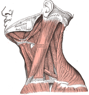
Mylohyoid muscle visible right under jaw
Image via Wikipedia

Do you notice symptoms of:

Or do any of these sound like you?

  1. Dry, itchy eyes or dry mouth
  2. Poor memory
  3. Unable to relax, become serene, or meditate
  4. History of diabetes in family
  5. Blood sugar problems, either hypoglycemia or diabetes
  6. Uncontrollable appetite (i.e., eating when not hungry)
  7. Desire to lose weight
  8. In need of a meal replacement

There is often a stress point involves the tongue, muscles of deglutination, and the posterior and lateral muscles of the neck. These muscles are involved with tongue movement, swallowing and the hyoid bone. When they are weak and contracted, hence the previous mentioned complaints.
#6/SvG – T1 to T3 – Stress Point Palpation

Other tissues that may be involved with this stress point are:

  • Retina
  • Muscles and tissues of the face and nose
  • Teeth

Effect of Carbohydrate Reduction in the Diet

The tissues prefer to use carbohydrate for energy over both protein and fat. The quantity of carbohydrate stores in the body is only a few hundred grams (glycogen in the liver and muscles.) This can supply energy needed for the body for about half a day. Therefore, after a few hours of carbohydrate deprivation the body resorts to converting protein and fat to glucose (in Stress Mode). Fat will be the primary source of energy production and depletion of its stores will continue unabated until depleted.

A very good product is SVG. It is essential to delivering the lacking carbohydrates.

Stretch Exercise
Practice the “tongue in cheek” movement. Hold for a count of five. Do each side at least three times several times daily.

Enhanced by Zemanta

Forward Head Posture and TMJ

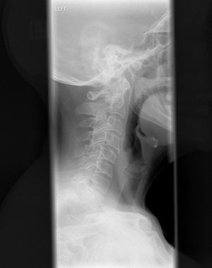
Xray of cervical spine
Image via Wikipedia

Forward head posture creates a domino effect in the body:

  • Moving forward, the head shifts the center of gravity.
  • To compensate, the upper body drifts backward.
  • To compensate for the upper body shift, the hips tilt forward.

So, the forward head position can be the cause of not only the head, neck and TMJ problems, but also mid back and low back problems. We can gain a better understanding of the interaction between the mouth opening and closing, TMJ bio-mechanics, and neck  function with an overview of the structures involved.

According to Rene Cailliet, Physical Medicine and Rehabilitation Director at U.S.C. states: “It’s an axiom … that the body follows the head … You can realign your entire body by moving your head … your head held in a forward position can pull your entire body out of line.” He goes on to explain that the vital lung capacity is reduced as much as 30%. The gastrointestinal system is affected, particularly the large intestine. When a hunched position is assumed, the body becomes rigid, and range of motion is affected. Since endorphin production is reduced, an increase in pain and discomfort results.

Another researcher Kapandji, in his classic text on spinal function states, “The anterior muscles of the neck … act as the long arm of a lever … they are powerful flexors of the head and cervical column … flattening the cervical column.”

Additionally, numerous investigators describe the effect of altered jaw position on head posture. Forward and side to side head position changes the jaw, throat, and tongue. It compresses the neck  joints causing muscular nerve entrapments. Nerve root compression or posterior neck vertebral joint irritation or restriction result in peripheral entrapment neuropathies. One common entrapment is the greater or lesser suboccipital nerves that pass between the occiput and atlas. This may cause headaches or refer pain to the facial region.

General Symptoms by Head Forward Posture problems
A. Chronically tired (lowered hemoglobin, immature cells) 89%
B. Increased nervous tension 86%
C. Malaise 61%
D. Restless sleep (awaken tired) 78%
E. Numbness in hands (awaken with arm or leg asleep) 32%
F. Cold hands and feet (poor circulation) 67%
G. Back aches and leg aches (tired, ache-all-over feeling) 47%
H. Thirsty (much water doesn’t satisfy) 43%
I. Restless nibbling at food (never satisfied)
J. Blood (variations in count and quality; many irregularly formed cells; cell walls even thickness one week after treatment)
K. Lowered hemoglobin
L. Lower thyroid activity (an almost constant finding)
M. Facial pallor
N. Dull, non-sparkling eyes

Forward head posture is an enormous problem of our society. Nearly 100% of new patients presenting in my office have it. If you have it and don’t correct it, it will lead to neck arthritis and damaged discs.

If you are interested in being checked for forward head posture or want more information visit www.nevadachiro.com.

Enhanced by Zemanta

FETAL ORIGINS: HOW LIFE IN THE WOMB SHAPES THE REST OF OUR LIVES

"Views of a Fetus in the Womb", Leon...
Image via Wikipedia

The connection between life in the womb and what happens later in life has been of great interest to mankind throughout history. But scientific evidence of any real connection was scarce until 1989, when a study found that low birth weight was a positive indicator for heart disease later in life. That was just the beginning.

Initial skepticism of the birth weight studies has given way to widespread acceptance, as newer studies confirmed the original. But new studies have also discovered exceptionally strong evidence that a stressful and/or toxic uterine environment directly affects not only the mental and physical health of babies, but can lead to a whole range of mental and physical problems that can crop up later in life.

These discoveries have spawned a whole new field of study, called “fetal origins”, which has convinced respected scientists, writers and environmentalists to call for serious changes in how we view – and care for – those critical nine months in everyone’s life called pregnancy.

Two recent books on the subject are getting a lot of favorable attention, and are definitely recommended reading for anyone considering pregnancy.

Science writer Annie Murphy Paul’s new 2010 book, Origins: How the Nine Months Before Birth Shape the Rest of Our Lives, was prompted by her personal decision to have a baby. The publisher says that Ms. Paul “interviews experts from around the world; discovers how individuals gestated during the Nazi siege of Holland in World War II were still feeling its consequences decades later; how pregnant women who experienced the 9/11 attacks passed their trauma on to their offspring in the womb; how a lab accident led to the discovery of a common household chemical that can harm the developing fetus; how the study of a century-old flu pandemic reveals the high personal and societal costs of poor prenatal experience.”

Another 2009 landmark book also discusses the same theme. More than Genes: What Science Can Tell Us About Toxic Chemicals, Development, and the Risk to Our Children. The author, Professor Dan Agin, a neuroscientist and molecular geneticist at the University of Chicago, lays out a powerful array of convincing evidence. Publisher’s Weekly says Agin’s book describes “a silent pandemic…causing untold damage to babies while they are in the womb. Toxic chemicals in the environment are assaulting developing fetuses, as are substances (such as alcohol and nicotine) ingested by pregnant women and are capable of dramatically altering developmental pathways. According to Agin, the role of the intrauterine environment has largely been ignored by scientists who look to genes and a child’s post-birth environment to explain behavior issues, mental illness and IQ.”

Fetal origins as a science may still be in its infancy, but we should not wait for more studies to “prove” what is already evident — pregnant women must be protected from trauma, stress, illness and known toxins like lead, dioxins, PCBs, radiation and pesticides. And we should regulate the tens of thousands of widely used chemicals that have never been tested for safety.

As New York Times columnist Nicholas D. Kristof said in a recent article on the subject, “…we have learned that a uterus is not a diving bell that insulates its occupant from the world’s perils.”

SOURCE: Simon & Schuster, 2010, http://books.simonandschuster.com/Origins/Annie-Murphy-Paul/9780743296625 Oxford University Press, 2009, http://www.oup.com/us/catalog/general/subject/Medicine/Genetics/?view=usa&ci=9780195381504 New York Times, http://www.nytimes.com/2010/10/03/opinion/03kristof.html

If you would like more information visit www.nevadachiro.com or call us at 565-7474.


Enhanced by Zemanta

Why Health Care Costs for LBP Are So High

Initiating care with an MD for back pain results in much higher health care costs than going to a DC, says study.

By Peter W. Crownfield

With the much-touted Choudhry/Milstein study already putting insurers and other health care stakeholders on notice that chiropractic care for back pain “is highly cost-effective [and] represents a good value in comparison to medical physician care and to widely accepted cost-effectiveness thresholds,” along comes “Cost of Care for Common Back Pain Conditions Initiated With Chiropractic Doctor vs. Medical Doctor / Doctor of Osteopathy as First Physician.”

Published in the December 2010 issue of JMPT, the study, a retrospective claims analysis of Blue Cross Blue Shield of Tennessee’s intermediate and large group fully insured population, determined that paid costs for episodes of care were 40 percent lower when care was initiated with a doctor of chiropractic compared to an allopathic provider. Even when risk-adjusting each patient’s costs to account for severity, paid costs for chiropractic patients were 20 percent lower than medical patients treated for low back pain.

“Our results support a growing body of evidence that chiropractic treatment of low back pain is less expensive than traditional medical care,” stated the study authors in their conclusion. “We found that episode cost of care for LBP initiated with a DC is less expensive than care initiated through an MD. … Our results suggest that insurance companies that restrict access to chiropractic care for LBP may, inadvertently, be paying more for care than they would if they removed these restrictions.”

Health Care Cost In their study, the researchers identified Blue Cross Blue Shield of Tennessee members with an LBP claim based on the presence of any of the following ICD-9 codes on a paid claim: 722 Intervertebral disk disorders, 724 Other and unspecified disorders of back, 729 Other disorders of soft tissues, 739 Nonallopathic lesions not elsewhere classified, 846 Sprains and strains of sacroiliac region, and 847 Sprains and strains of other and unspecified parts of back. Of more than 650,000 members during the two-year period analyzed (Oct. 1, 2004 – Sept. 30, 2006), 85,402 had been diagnosed using one of the above codes.

Plan members had open access to MDs and DCs through self-referral (ER visits were categorized as MD-initiated care), without any limit to the number of visits. Co-pays did not vary between provider type.

Total episode costs for each episode of LBP were determined by calculating the cost paid by the insurer for all services provided during the episode by the same and other providers. Costs per episode were $452.33 (paid) for patients initiating care with a chiropractor and $1,037.04 for patients initiating care with a medical provider; risk-adjusted paid costs were $532.54 (DC) vs. $661.10 (MD).

“As doctors of chiropractic, we know firsthand that our care often helps patients avoid or reduce more costly interventions such as drugs and surgery. This study supports what we see in our practices every day,” said ACA President Rick McMichael, DC, in an ACA release reporting on the study findings. “It also demonstrates the value of chiropractic care at a critical time, when our nation is attempting to reform its health care system and contain runaway costs.”

Enhanced by Zemanta